Main R&D areas
Bioinformatics and Systems Biology Lab | BioSys_BUT
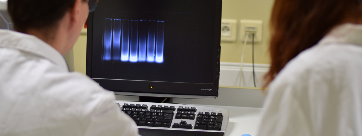
Our research team develops new computational tools for the analysis of omics data to perform rapid typing of bacteria, gene regulatory networks inference, or non-coding elements detection. We also focus on advanced and holistic analysis of microbial organisms, especially bacteria. In addition to pathogenic bacteria from the hospital environment associated with nosocomial infections, we focus on non-model species with biotechnological potential, particularly bioplastics or biofuel producers. Equally important areas of our research are the analysis of microbial communities, for example, those from the gut of farm animals, and the investigation of the relationship between biological activity and the structure of organic substances. We work intensively in cooperation with clinical experts from the University Hospital Brno, biotechnologists from the Faculty of Chemistry BUT, colleagues from Masaryk University, the Czech Collection of Microorganisms and the Veterinary Research Institute and several foreign workplaces, for example, LMU in Munich or UNL in the USA.
Lab of Bioengineering and Applied Biomaterials | BioEngiMat_BUT
Research group focuses on bio-materials in a broad sense, from the development of innovative bio-materials to the understanding of their interaction with cells to the conception of preparation of scaffolds, implants or 2D in vitro platforms with desired properties. Researchers use the principles of both engineering and life sciences including 3D (bio)printing, patterning or electrospray deposition method and basic methods in cellular and molecular biology. The group also deals with the synthesis and modification of nanomaterials with antibacterial properties, bioactivity, and as the filler improving mechanical/electrical properties of hydrogels and scaffolds.
Biomedical Signal Processing Group | BioSig_BUT
We measure and analyze biological signals. Our main focus is the measurement and processing of both clinical and experimental cardiological data (electrocardiograms, photoplethysmograms). We also focus on the processing and analysis of data from wearable devices (smartphones, watches, and bands) to assess individuals' health status and activity levels. Additionally, we deal with the processing of multimodal data, such as for detecting sleep apnea, analyzing psychological conditions, detecting stress in drivers and soldiers, or evaluating athletes' performance. We collaborate closely with doctors and psychologists from Masaryk University, the Center for Sports Activities at Brno University of Technology, ICRC, Brno University Hospital, the Institute of Instrumentation of the Academy of Sciences of the Czech Republic, Mayo Clinic, and the Office of Naval Research.
Biomedical imaging and image analysis | BioImage_BUT
We develop advanced software tools for various biomedical images, enabling detailed analysis and processing. The group focuses on developing complex algorithms for the analysis and processing of medical imaging data, such as CT and MR, to support medical diagnostics. Additionally, we specialize in capturing and analyzing ophthalmological data, particularly retinal images, and in analyzing images from microscopic techniques. Our activities include theoretical research, the creation of new algorithms, their implementation, and clinical testing in collaboration with major international and national institutions.
